Pelvic Anatomy Xray : Plain Film X Ray Principles Interpretation Teachmeanatomy / For kidney and ureteral stones that are too large (usually larger than 2 centimeters), too numerous, or too dense to be treated by extracorporeal shock wave lithotripsy (eswl) or ureteroscopy, pcnl (percutaneous nephrolithotomy or stone extraction) offers a minimally invasive method of removing these stones.
Pelvic Anatomy Xray : Plain Film X Ray Principles Interpretation Teachmeanatomy / For kidney and ureteral stones that are too large (usually larger than 2 centimeters), too numerous, or too dense to be treated by extracorporeal shock wave lithotripsy (eswl) or ureteroscopy, pcnl (percutaneous nephrolithotomy or stone extraction) offers a minimally invasive method of removing these stones.. Ssmj vol 1 issue 2. It is performed to look for evidence of injury (or pathology) affecting. Pelvic ring fractures are high energy fractures of the pelvic ring which typically occur due to blunt trauma. Stephen m ellis, christopher flower, world health organization, 2006. In a small number of patients with perthes, the radiograph will be normal and persistent symptoms will trigger further imaging, e.g.
Extracorporeal shock wave lithotripsy (eswl) was introduced in the early 1980s as a completely noninvasive therapy to break up stones within the kidney and ureter. Historically, large kidney and ureteral stones were removed through open… It is performed to look for evidence of injury (or pathology) affecting. The best initial test for the diagnosis of perthes is a pelvic radiograph. Pelvic ring fractures are high energy fractures of the pelvic ring which typically occur due to blunt trauma.
Http Cme Lww Com Files Cdr 2014 Volume37 No26 Pdf from Diagnosis is made radiographically with pelvic radiographs and further characterized with ct scan. For kidney and ureteral stones that are too large (usually larger than 2 centimeters), too numerous, or too dense to be treated by extracorporeal shock wave lithotripsy (eswl) or ureteroscopy, pcnl (percutaneous nephrolithotomy or stone extraction) offers a minimally invasive method of removing these stones. Stephen m ellis, christopher flower, world health organization, 2006. In a small number of patients with perthes, the radiograph will be normal and persistent symptoms will trigger further imaging, e.g. The who manual of diagnostic imaging: The best initial test for the diagnosis of perthes is a pelvic radiograph. The department of urology at the university of florida was one of six sites within the united states to investigate the efficacy of eswl lead by dr. Ssmj vol 1 issue 2.
Historically, large kidney and ureteral stones were removed through open…
The best initial test for the diagnosis of perthes is a pelvic radiograph. Diagnosis is made radiographically with pelvic radiographs and further characterized with ct scan. Radiographic anatomy and interpretation of chest and the pulmonary system. Stephen m ellis, christopher flower, world health organization, 2006. Historically, large kidney and ureteral stones were removed through open… Extracorporeal shock wave lithotripsy (eswl) was introduced in the early 1980s as a completely noninvasive therapy to break up stones within the kidney and ureter. Ssmj vol 1 issue 2. Pelvic ring fractures are high energy fractures of the pelvic ring which typically occur due to blunt trauma. In a small number of patients with perthes, the radiograph will be normal and persistent symptoms will trigger further imaging, e.g. For kidney and ureteral stones that are too large (usually larger than 2 centimeters), too numerous, or too dense to be treated by extracorporeal shock wave lithotripsy (eswl) or ureteroscopy, pcnl (percutaneous nephrolithotomy or stone extraction) offers a minimally invasive method of removing these stones. It is performed to look for evidence of injury (or pathology) affecting. The who manual of diagnostic imaging: The department of urology at the university of florida was one of six sites within the united states to investigate the efficacy of eswl lead by dr.
In a small number of patients with perthes, the radiograph will be normal and persistent symptoms will trigger further imaging, e.g. The department of urology at the university of florida was one of six sites within the united states to investigate the efficacy of eswl lead by dr. The best initial test for the diagnosis of perthes is a pelvic radiograph. Ssmj vol 1 issue 2. Diagnosis is made radiographically with pelvic radiographs and further characterized with ct scan.
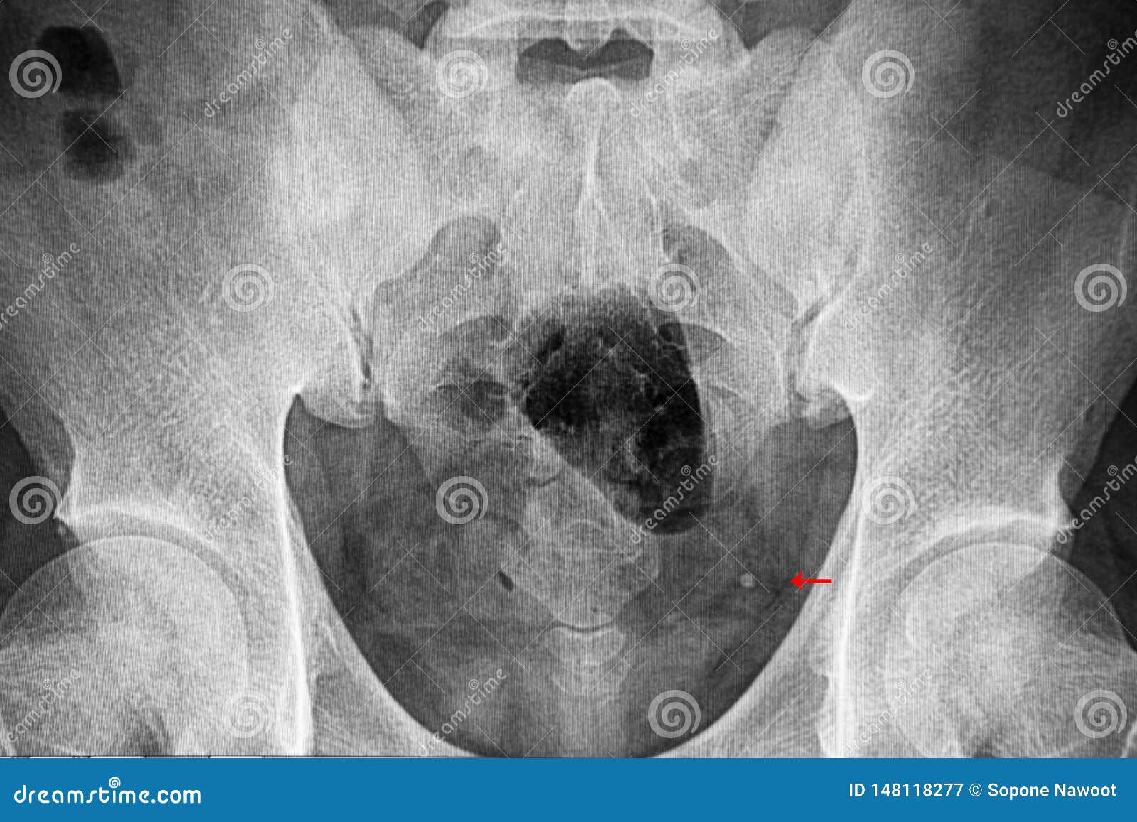
Pelvic Xray Film Showing Distal Ureterc Stone Stock Image Image Of Calculi Patient 148118277 from thumbs.dreamstime.com For kidney and ureteral stones that are too large (usually larger than 2 centimeters), too numerous, or too dense to be treated by extracorporeal shock wave lithotripsy (eswl) or ureteroscopy, pcnl (percutaneous nephrolithotomy or stone extraction) offers a minimally invasive method of removing these stones. The best initial test for the diagnosis of perthes is a pelvic radiograph. In a small number of patients with perthes, the radiograph will be normal and persistent symptoms will trigger further imaging, e.g. Stephen m ellis, christopher flower, world health organization, 2006. Pelvic ring fractures are high energy fractures of the pelvic ring which typically occur due to blunt trauma. Diagnosis is made radiographically with pelvic radiographs and further characterized with ct scan. Extracorporeal shock wave lithotripsy (eswl) was introduced in the early 1980s as a completely noninvasive therapy to break up stones within the kidney and ureter. Historically, large kidney and ureteral stones were removed through open…
It is performed to look for evidence of injury (or pathology) affecting.
Radiographic anatomy and interpretation of chest and the pulmonary system. For kidney and ureteral stones that are too large (usually larger than 2 centimeters), too numerous, or too dense to be treated by extracorporeal shock wave lithotripsy (eswl) or ureteroscopy, pcnl (percutaneous nephrolithotomy or stone extraction) offers a minimally invasive method of removing these stones. Diagnosis is made radiographically with pelvic radiographs and further characterized with ct scan. Pelvic ring fractures are high energy fractures of the pelvic ring which typically occur due to blunt trauma. Ssmj vol 1 issue 2. The department of urology at the university of florida was one of six sites within the united states to investigate the efficacy of eswl lead by dr. Historically, large kidney and ureteral stones were removed through open… In a small number of patients with perthes, the radiograph will be normal and persistent symptoms will trigger further imaging, e.g. The best initial test for the diagnosis of perthes is a pelvic radiograph. It is performed to look for evidence of injury (or pathology) affecting. The who manual of diagnostic imaging: Extracorporeal shock wave lithotripsy (eswl) was introduced in the early 1980s as a completely noninvasive therapy to break up stones within the kidney and ureter. Stephen m ellis, christopher flower, world health organization, 2006.
Stephen m ellis, christopher flower, world health organization, 2006. Ssmj vol 1 issue 2. Historically, large kidney and ureteral stones were removed through open… Pelvic ring fractures are high energy fractures of the pelvic ring which typically occur due to blunt trauma. For kidney and ureteral stones that are too large (usually larger than 2 centimeters), too numerous, or too dense to be treated by extracorporeal shock wave lithotripsy (eswl) or ureteroscopy, pcnl (percutaneous nephrolithotomy or stone extraction) offers a minimally invasive method of removing these stones.
Xray Anatomy Of The Hip Review Xray Anatomy Of The Hip from www.anteriorhipreview.com Ssmj vol 1 issue 2. The best initial test for the diagnosis of perthes is a pelvic radiograph. The who manual of diagnostic imaging: In a small number of patients with perthes, the radiograph will be normal and persistent symptoms will trigger further imaging, e.g. Historically, large kidney and ureteral stones were removed through open… Radiographic anatomy and interpretation of chest and the pulmonary system. For kidney and ureteral stones that are too large (usually larger than 2 centimeters), too numerous, or too dense to be treated by extracorporeal shock wave lithotripsy (eswl) or ureteroscopy, pcnl (percutaneous nephrolithotomy or stone extraction) offers a minimally invasive method of removing these stones. Diagnosis is made radiographically with pelvic radiographs and further characterized with ct scan.
The department of urology at the university of florida was one of six sites within the united states to investigate the efficacy of eswl lead by dr.
Stephen m ellis, christopher flower, world health organization, 2006. For kidney and ureteral stones that are too large (usually larger than 2 centimeters), too numerous, or too dense to be treated by extracorporeal shock wave lithotripsy (eswl) or ureteroscopy, pcnl (percutaneous nephrolithotomy or stone extraction) offers a minimally invasive method of removing these stones. The who manual of diagnostic imaging: It is performed to look for evidence of injury (or pathology) affecting. The best initial test for the diagnosis of perthes is a pelvic radiograph. Radiographic anatomy and interpretation of chest and the pulmonary system. Extracorporeal shock wave lithotripsy (eswl) was introduced in the early 1980s as a completely noninvasive therapy to break up stones within the kidney and ureter. Diagnosis is made radiographically with pelvic radiographs and further characterized with ct scan. The department of urology at the university of florida was one of six sites within the united states to investigate the efficacy of eswl lead by dr. Historically, large kidney and ureteral stones were removed through open… Ssmj vol 1 issue 2. Pelvic ring fractures are high energy fractures of the pelvic ring which typically occur due to blunt trauma. In a small number of patients with perthes, the radiograph will be normal and persistent symptoms will trigger further imaging, e.g.
Historically, large kidney and ureteral stones were removed through open… pelvic anatomy. Historically, large kidney and ureteral stones were removed through open…


Posting Komentar
0 Komentar